Digital Radiography and CT-Scan Findings in Femoral Bone Structures of Long-legged Buzzard (Buteo rufinus)
Keywords:
Long-legged buzzard, Femur, Digital radiology, CT-scan, Buteo rufinusAbstract
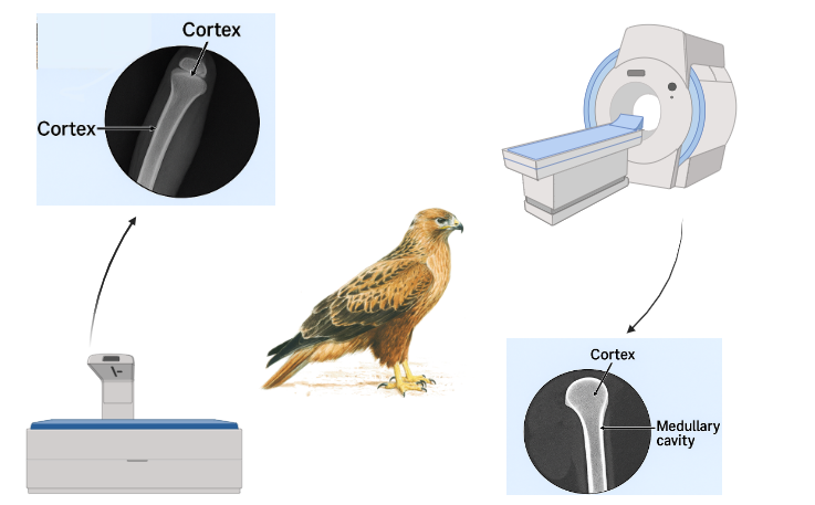
Veterinary diagnostic methods are rapidly evolving, with radiology techniques playing a key role in clinical activities and disease diagnosis. Computed Tomography (CT) has gained popularity for its detailed structural imaging and ability to provide precise diagnostic information. The Buteo rufinus, a common bird of prey species admitted to the Environmental Protection Department of Kerman, often presents with skeletal injuries, making advanced imaging techniques valuable for effective treatment. This study examined the femoral bone structure using digital radiology and CT in 10 adult Buteo Rufinus. Sedated birds underwent imaging of left and right femur structures, and multiple indices were measured. A significant difference was found in the diameter of the medullary cavity and thickness of the cortex between radiology and CT techniques (P<0.05), while no significant difference was observed between left and right femoral indices using either method (P>0.05). CT offers higher accuracy in measuring some femur indices and provides precise information for managing femoral injuries in Buteo rufinus. It is recommended for enhancing clinical outcomes in these raptors
Downloads
References
Khaleghizadeh A, Mohammad K, Mansour A, Mohammad T, Alireza H, Seyed Babak M, et al. Atlas of Birds of Iran: Iran Department of the Environment, Tehran; 2020. 617-103 p.
Forbes NA. Avian orthopedics. Veterinary Quarterly. 1998;20(sup1):S69-70. doi: 10.1080/01652176.1998.10807421.
Molina-López RA, Casal J, Darwich L. Causes of morbidity in wild raptor populations admitted at a wildlife rehabilitation centre in Spain from 1995-2007: a long term retrospective study. PLoS One. 2011;6(9):e24603. doi: 10.1371/journal.pone.0024603.
Silva IA, Vieira LC, Mancini VRM, Faillace ACL, Santana MIS. Radiographic anatomy of the cockatiel (Nymphicus hollandicus) axial and appendicular skeleton. Anat Histol Embryol. 2020;49(2):184-95. doi: 10.1111/ahe.12510.
Naguib M. Avian radiography and radiology part 2. Companion Anim. 2017;22(10):614-21. doi: 10.12968/coan.2017.22.10.614.
Bennett RA, Kuzma AB. Fracture management in birds. Journal of Zoo and Wildlife Medicine. 1992:5-38.
Orsini JA, Grenager NS, de Lahunta A. Comparative Veterinary Anatomy: A Clinical Approach: Academic Press; 2021.
Ricciardi M, Franchini D, Valastro C, Ciccarelli S, Caprio F, Eyad Assad A, et al. Multidetector computed tomographic anatomy of the lungs in the loggerhead sea turtle (Caretta caretta). Anat Rec. 2019;302(9):1658-65. doi: 10.1002/ar.24030.
Hanna AL, Logsdon ML, Mattoon JS. Radiographic Evaluation of Normal and Common Diseases2021. 173-91 p.
Krautwald-Junghanns ME, Kostka VM, Dörsch B. Comparative studies on the diagnostic value of conventional radiography and computed tomography in evaluating the heads of psittacine and raptorial birds. J Avian Med Surg. 1998:149-57.
Nejad MRE, Vafaei R, Masoudifard M, Nassiri SM, Salimi A. Aggressive chondroblastic osteosarcoma in a dog: A case report: Faculty of Veterinary Medicine, Urmia University, Urmia, Iran; 2019. 361 p.
Delibaş V, Soygüder Z, Göya C, Aslan L, Çakmak G. Three-Dimensional Examination of Humerus and Antebrachium Bones in the Red hawk (Buteo Rufinus) with Computed tomography (CT). Van Veterinary Journal. 2024;35(1):70-6. doi: 10.36483/vanvetj.1396960.
Lautenschlager S, Bright JA, Rayfield EJ. Digital dissection-using contrast‐enhanced computed tomography scanning to elucidate hard‐and soft‐tissue anatomy in the common buzzard Buteo buteo. J Anat. 2014;224(4):412-31. doi: 10.1111/joa.12153.
Habib MB, Ruff CB. The effects of locomotion on the structural characteristics of avian limb bones. Zool J Linn Soc. 2008;153(3):601-24. doi: 10.1111/j.1096-3642.2008.00402.x.
Bertuccelli T, Crosta L, Costa GL, Schnitzer P, Sawmy S, Spadola F. Predisposing anatomical factors of humeral fractures in birds of prey: a preliminary tomographic comparative study. J Avian Med Surg. 2021;35(2):123-34. doi: 10.1647/19-00006.
de Lima LFS, de Barros AJBP, Colodel EM, Nespoli PEB, Gomes LG, de Souza RL. Radiographic, tomography and three-dimensional description of the clinical anatomy of the long bones of Rupornis Magnirostris. OBSERVATÓRIO DE LA ECONOMÍA LATINOAMERICANA. 2023;21(10):16852-73. doi: 10.55905/oelv21n10-126.
Scholarship W, Schneider MA. Birds of the ancient Nile: Species identification in Egyptian animal mummies using multi-resolution computed tomography and deep learning image segmentation 2024.
Fajardo RJ, Hernandez E, O'Connor PM. Postcranial skeletal pneumaticity: a case study in the use of quantitative microCT to assess vertebral structure in birds. J Anat. 2007;211(1):138-47. doi: 10.1111/j.1469-7580.2007.00749.x.
Burghardt AJ, Link TM, Majumdar S. High-resolution computed tomography for clinical imaging of bone microarchitecture. Clin Orthop Relat Res. 2011;469(8):2179-93. doi: 10.1007/s11999-010-1766-x.
Orosz S, Echols S, Redig P. Avian Surgical Anatomy and Orthopedic Management: Teton NewMedia; 2023.

Downloads
Published
Issue
Section
License
Copyright (c) 2024 Basir Abolghasemi (Author); Mohammadreza Esmailinejad (Corresponding Author); Hemad Shafiei, Mohammadjavad Jahedi, Nafiseh Alinejad (Author)

This work is licensed under a Creative Commons Attribution-NonCommercial 4.0 International License.