Localized Non-Pigmented Villonodular Synovial Proliferation in a Domestic Canary (Serinus canaria domestica): A Case Report and Pathological Findings
Keywords:
Villonodular synovitis, Canary (Serinus canaria), Avian pathology, Synovial proliferation, Joint diseaseAbstract
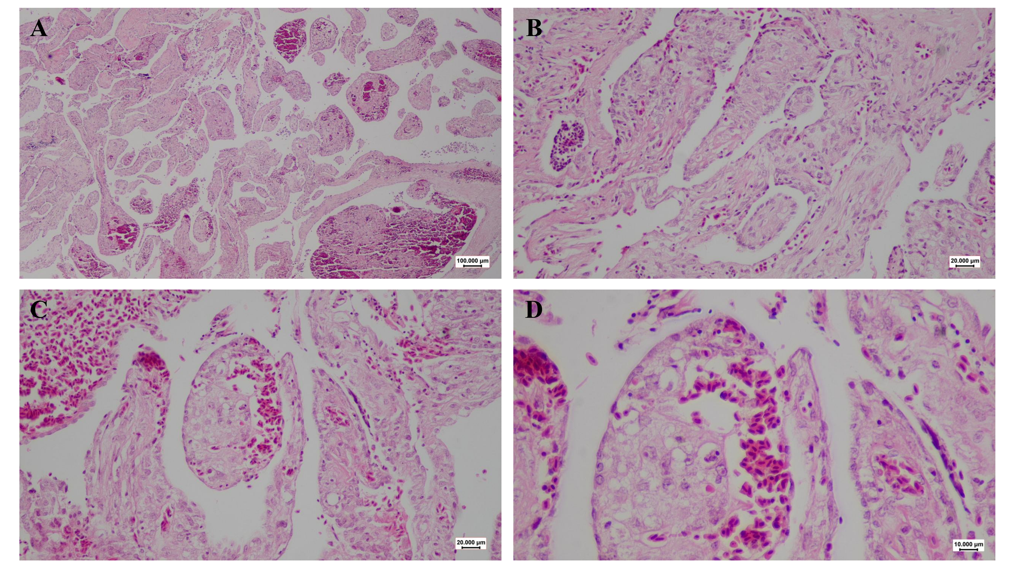
Villonodular synovitis (VNS) is a rare, proliferative disorder of the synovial membrane that occurs predominantly in mammals, particularly humans and large domestic animals. It presents as localized or diffuse forms characterized by villous and nodular synovial hyperplasia. While VNS is well documented in human and veterinary orthopaedic literature, reports in birds are exceptionally rare. An adult domestic canary (Serinus canaria domestica) was presented with a dome-shaped nodular swelling at the left wing. The bird was maintained under indoor breeding conditions with a seed-based diet. Surgical excision was performed under isoflurane anaesthesia following standard aseptic preparation. The mass was removed via blunt and sharp dissection, and the wound was closed in two layers. Postoperative care included meloxicam and enrofloxacin administration, with uneventful recovery and complete wound healing within two weeks. Histopathological examination revealed a localized papillary, villous, and nodular synovial-type proliferation supported by fibrovascular stroma, with synovial-lined cleft-like spaces, and without detectable hemosiderin-laden macrophages or multinucleated giant cells. The findings were most consistent with a localized non-pigmented VNS-like synovial proliferative lesion. The lesion's localization, absence of pigment, and uneventful short-term postoperative recovery suggest a favourable immediate outcome. This report highlights the need to include synovial proliferative disorders in the differential diagnosis of avian joint swellings and underscores the diagnostic value of histopathology in such cases.
Downloads

Published
Issue
Section
License
Copyright (c) 2026 Shahrzad Azizi, Mohammad Farajli Abbasi, Aysel Mahalati, Reza Kheirandish (Author); Sina Delshad (Corresponding Author)

This work is licensed under a Creative Commons Attribution-NonCommercial 4.0 International License.